Fig 1.

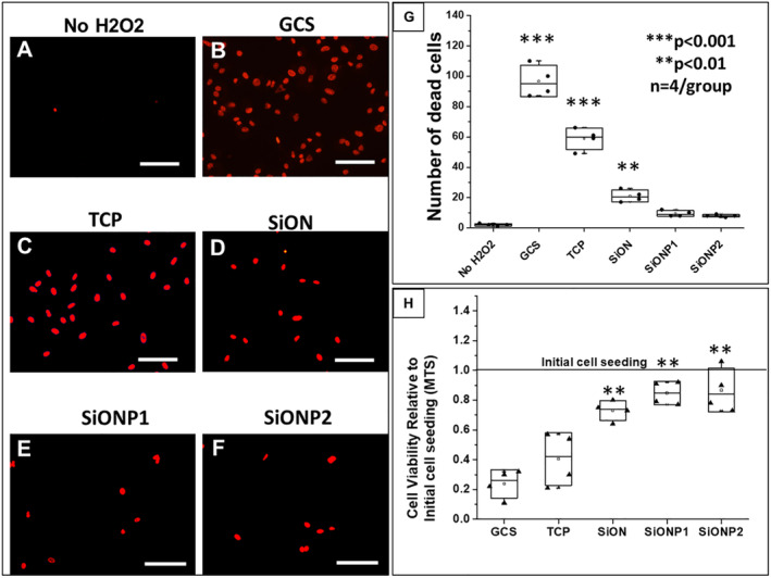
Effect of silicon oxynitride‐ (SiONx‐) and silicon oxynitrophosphide‐ (SiONPx‐) plasma‐enhanced chemical vapor deposition nanoscale implant coating on human umbilical vein endothelial cells under toxic levels of hydrogen peroxide (H2O2; 24 hours). Scale bar = 100 μm. (A–F) Propidium iodide (PI) staining shows dead cells (red stain). (G) Analysis after ANOVA (Tukey's pairwise) shows data from PI counting according to group. (H) Comparison of cell viability relative to the initial cell seeding among groups after MTS assay, the data were analyzed by ANOVA (Tukey's pairwise). GCS = glass cover slip; SiONP = silicon oxynitrophosphide; TCP = tissue culture plate. n = 4. ***p < 0.001 and **p < 0.01, all compared with No H2O2 group.
