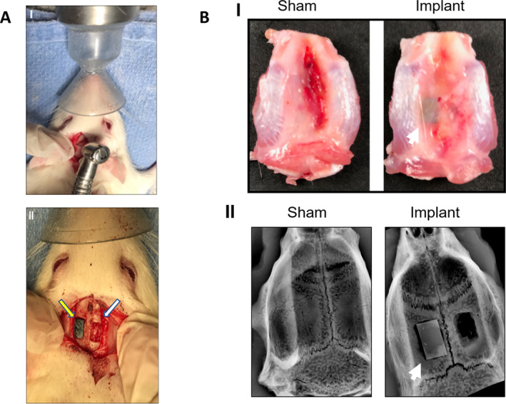
Fig 6.

(A) Surgical procedure for material implantation. (I.) Gross image of calvarial defect surgery with dental bur. (II) Gross image of the parietal bone bilateral calvarial defect (6 × 4 mm), implant on the left (yellow arrow) and empty on the right (white arrow). (B) Samples harvested from rat calvarium 15 days after surgery. (I) Gross image shows the macroscopic superior aspect of the calvaria. (II.) An X‐ray image with the sham (left) and calvarial defects and implant (right). The white arrow points to the implant. n = 3.
