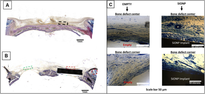
Fig 8.

Bright field images acquired using the BIOQUANT Osteoimager showing coronal sections of rat calvaria after Sanderson's staining. Dotted rectangles represent the areas used for capturing immunofluorescent images. (A) The dotted black rectangular box is representative of a sham‐operated animal. (B) The dotted green rectangular box indicates the location assessed for the empty standard‐size calvarial defect, while the dotted red rectangular box indicates the location of the assessed implant‐filled standard‐size calvarial defect. The muscle is traced with a red circle. Scale bar = 1 mm. (C) Higher magnification images of empty defect (showing no vascular tissue formation) and coated implant surfaces (showing new vascular tissue formation). Uncoated surfaces did not exhibit any new vascular tissue formation, which matched that of empty defects. SiONP = silicon oxynitrophosphide.
