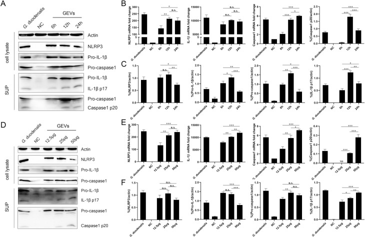
Fig 5. Activation of NLRP3 inflammasome in murine peritoneal macrophages.
Cells were incubated with 25 μg/mL of GEVs for 6 h, 12 h and 24 h or incubated for 12 h with 12.5, 25, 50 μg/mL of GEVs. Protein expression of NLRP3 (110 KDa), Pro-IL-1β (35 KDa), Pro-caspase1(45 KDa) in the cell lysate and IL-1β p17 (17 KDa), Caspase1 p20 (20 KDa) in the SUP were detected using western blot (A and D). The mRNA fold changes of NLRP3, IL-1β, Caspase1 were detected using qPCR (B and E). The protein expression levels were measured by calculating the ratio percentage of band density in target protein and housekeeping actin. (C and F). NC represented No GEVs-treated negative control group. G. duodenalis in A represented positive control with 1.5 × 10⁶ parasites/mL for 24 h and in D represented positive control with 1.5 × 10⁶ parasites/mL for 12 h. SUP represented cell culturing supernatants. The data are expressed as the mean ± SEM from three separate experiments, *p < 0.05, **p < 0.01 or ***p <0.001 for different treated time groups or different treated amount groups.

