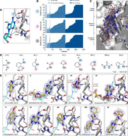
Fig. 4. Fragments binding to the adenine subsite.

(A) Stick representation showing the interaction of the adenosine moiety of ADPr with Mac1. The key hydrogen bonds are shown as dashed lines. (B) Plot of the distances shown in (A) for all fragment hits. The distances, truncated to 10 Å, are for the closest noncarbon fragment atom. (C) Stick representation showing all fragments interacting with Asp22-N, Ile23-N, or Ala154-O. The surface is “sliced” down a plane passing through Asp22. (D) Structures of the nine unique motifs that make at least two hydrogen bonds to the adenine subsite. Colored circles match the interactions listed in (A) and (B). The number of fragments identified for each motif are listed in parentheses. (E) Examples of the nine structural motifs. The fragment is shown with yellow sticks and the PanDDA event map is shown as a blue mesh. ADPr is shown as cyan transparent sticks. The apo structure is shown with dark gray transparent sticks.
