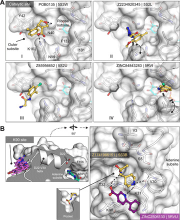
Fig. 6. Fragments targeting the catalytic and potential allosteric sites are sparsely populated compared to the adenosine site.

(A) Surface representation showing fragments that bind near the catalytic site. The fragment POB0135 (PDB 5S3W) bridges the gap between Asn40 and Lys102 via a hydrogen bond and a salt bridge, respectively. Although eight fragments bind in the outer subsite, the fragment POB0135 makes the highest-quality interactions. No fragments bind in the ribose subsite. The fragment ZINC331715 (PDB 5RVI) inserts into the phosphate subsite between Ile131 and Gly47. (B) Left: The K90 site is connected to the adenosine site by the Asp22-Val30 α helix. Right: Surface representation showing two fragments that bind to the K90 site. Hydrogen bonds are shown as dashed black lines. The fragment Z1741966151 (PDB 5S3B) is partially inserted in a nearby pocket (inset).
