Figure 3.

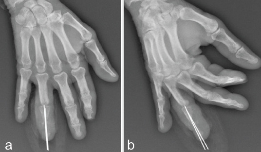
(a and b) Radiographs (anteroposterior and lateral view) showing extensive osteomyelitis of the middle and distal phalanx with significant bone lucency and osteopenia.

(a and b) Radiographs (anteroposterior and lateral view) showing extensive osteomyelitis of the middle and distal phalanx with significant bone lucency and osteopenia.