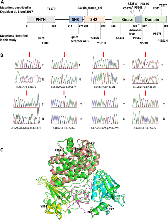
Figure 1: Follicular lymphoma-associated BTK mutations are distributed over the BTK coding region and cluster on the protein surface.

A: Location of BTK mutations identified by Krysiak et al Blood 2017 and in this study in a linear schema of BTK (22). B: Sanger sequence traces of paired sorted FL B cell (T) and paired CD3 T cell (N) DNA. Mutated residues are indicated by a red arrow. C: Composite 3-D model of BTK with protein domains and location of identified BTK missense mutations.
