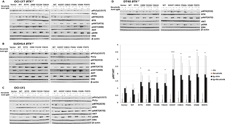
Figure 4: BTK mutations enhance anti-IG induced AKT phosphorylation.

A–D: Engineered OCI-LY7 BTK−/−, SUDHL4 BTK−/−, or DT40 BTK−/− cell lines as well as OCI-LY1 cells expressing low level endogenous BTK were reconstituted with HA-tagged BTK WT or mutant cDNAs and stable cell lines generated. The cells were rested for 1 h in serum-free medium and cell aliquots pre-treated for 1 h with 1 µM of ibrutinib followed by stimulation for 10’ with 10 µg / ml of anti-IG antibodies. Detergent cell lysates were made, fractioned by SDS-PAGE and prepared for immunoblotting with antibodies targeting the indicated epitopes. Displayed are immunoblotting data from representative experiments (vector: cells infected with empty FG9 lentivirus). Displayed are composite densitometry data from three independent experiments per cell line for the p-AKT473:AKT data (paired two-sample t-test; * p<0.05; ** p<0.01; *** p<0.001).
