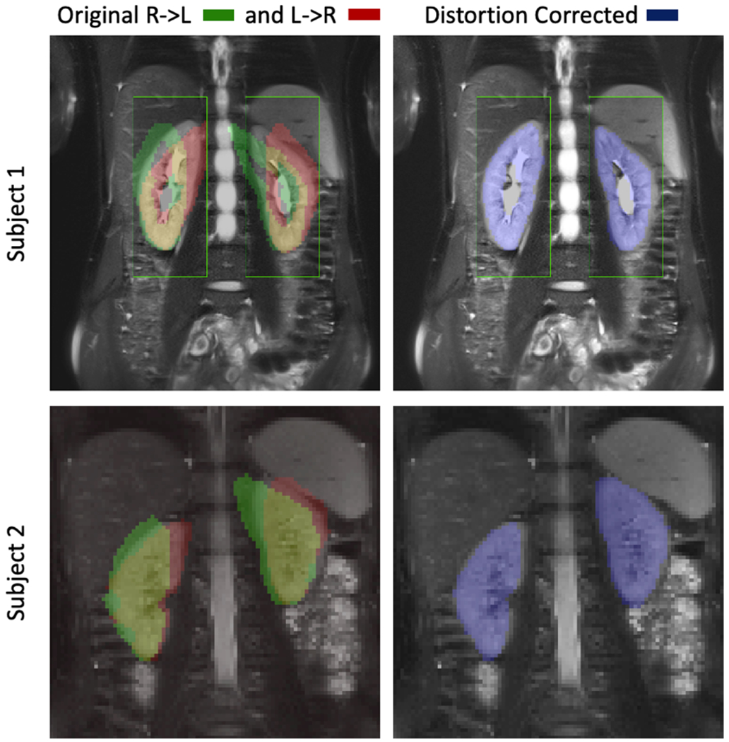
Figure 2.

Reference T2-HASTE image and the segmented kidney masks from the DW images are shown for the L->R and R->L images without correction on the left and for the distortion corrected image on the right. Each row corresponds to one representative subject. The kidneys are severely distorted in the original DW images. After distortion correction, the kidneys are in good alignment with the reference image, which resulted in an increase in Likert score from 2.6+/−1.0 to 3.7+/−1.0 (p<0.05).
