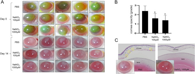
Figure 9.
In vivo effect of NO on corneal opacity development after alkali burn. (A) Representative photographs of the alkali burned murine cornea at day 0 and 14 days after injury. Green stained areas by fluorescein at day 0 represent corneal epithelial defect induced by alkali burn. Corneal opacity of NO treated corneas (100 μM and 1000 μM) was significantly better than PBS control observed at 2 weeks. (B) The difference of corneal opacity grades between treatments and control (PBS) was significant. However, the difference between 100 and 1000 μM of sodium nitrite treatments was not significant. (C) Histologic examination revealed significantly increased corneal thickness and stromal cellularity (yellow arrows) in PBS treated cornea compared to NO treated cornea. *p < 0.05, scale bar: 100 μm.

