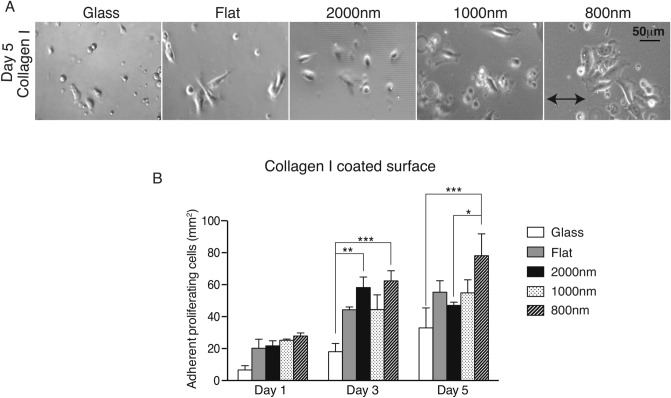
Figure 3.
Combination of collagen I and smaller topography size resulted in improving cellular spreading and growth. (A) MCEC were seeded on collagen I coated glass and 4 different silk topographies; flat, 2000 nm, 1000 nm and 800 nm ridge width pattern. Phase-contrast images were taken at 5-day time point. (B) Quantification of cell number demonstrated that collagen I coating improved cell spreading and growth when compared to previous experiments. Silk films were better than glass and the smallest ridge width 800 nm showed the highest cell number (* indicates p < 0.05, ** indicates p < 0.005, ***indicates p < 0.0005. Values are means ± S.E.M. The arrow indicates the direction of pattern axis).

