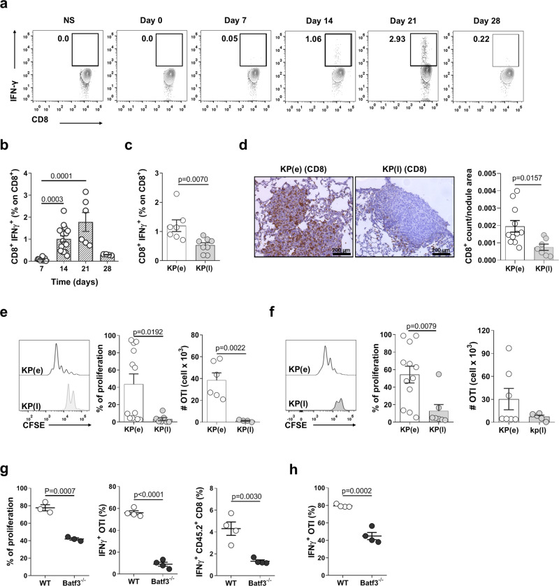
Fig. 2. CD8+ T cell responses and cross-presentation of tumor antigens by cDC1 are inhibited in late tumors.
a, b Representative plots and quantification of the kinetic of IFN-γ production by endogenous blood CD8+ T cells in KP-OVA challenged mice. Data are from two independent experiments at day 7–28 or 3 independent experiments at day 14–21. In b Kruskal–Wallis followed by Dunn’s post-test day 7 n = 6, day 14 n = 15, day 21 n = 6, day 28 n = 4 mice. c Percentage of IFN-γ producing endogenous CD8+ T cells in lungs bearing early or late KP-OVA tumors. KP(e) n = 7, KP(l) n = 8 mice in two independent experiments, two-tailed unpaired t-test. d CD8+ T cells localization in early and late KP-OVA tumors. Scale bar 200 µm. Quantification of the density of CD8+ T cells as a function of the nodule area was calculated automatically on three consecutive sections/sample. Data are from KP(e) n = 11, KP(l) n = 7, two-tailed unpaired t-test. e, f Proliferation profile of OT-I in the MLN (e) or lung (f) of mice bearing early or late KP-OVA tumors was measured 2 days after transfer. Proliferation index (cells that underwent at least two cycles of proliferation) and absolute numbers of proliferated cells are depicted. In e % of proliferation KP(e) n = 12, KP(l) n = 7; absolute numbers KP(e) n = 6, KP(l) n = 4. In f %proliferation KP(e) n = 12, KP(l) n = 7; absolute numbers KP(e) n = 7, KP(l) n = 5. Data are from three independent experiments, two-tailed unpaired t-test. g, h KP-OVA tumors were induced in WT and Batf3−/− mice. g Proliferation and IFN-γ production of adoptively transferred OVA specific CD8 T cells and IFN-γ production by endogenous CD8+ T cells in the lung. %proliferation WT n = 3, Batf3−/− n = 3; IFNγ+OTI (%) and IFNγ+CD45.2+CD8(%) WT n = 4, Batf3−/− n = 4; two-tailed unpaired t-test. h IFN-γ production by OT-I in MLNs. Data represent one of two independent experiments (n = 4 mice/group), two-tailed unpaired t-test. All data are expressed as mean ± SEM. Source data are provided as a Source Data file.

