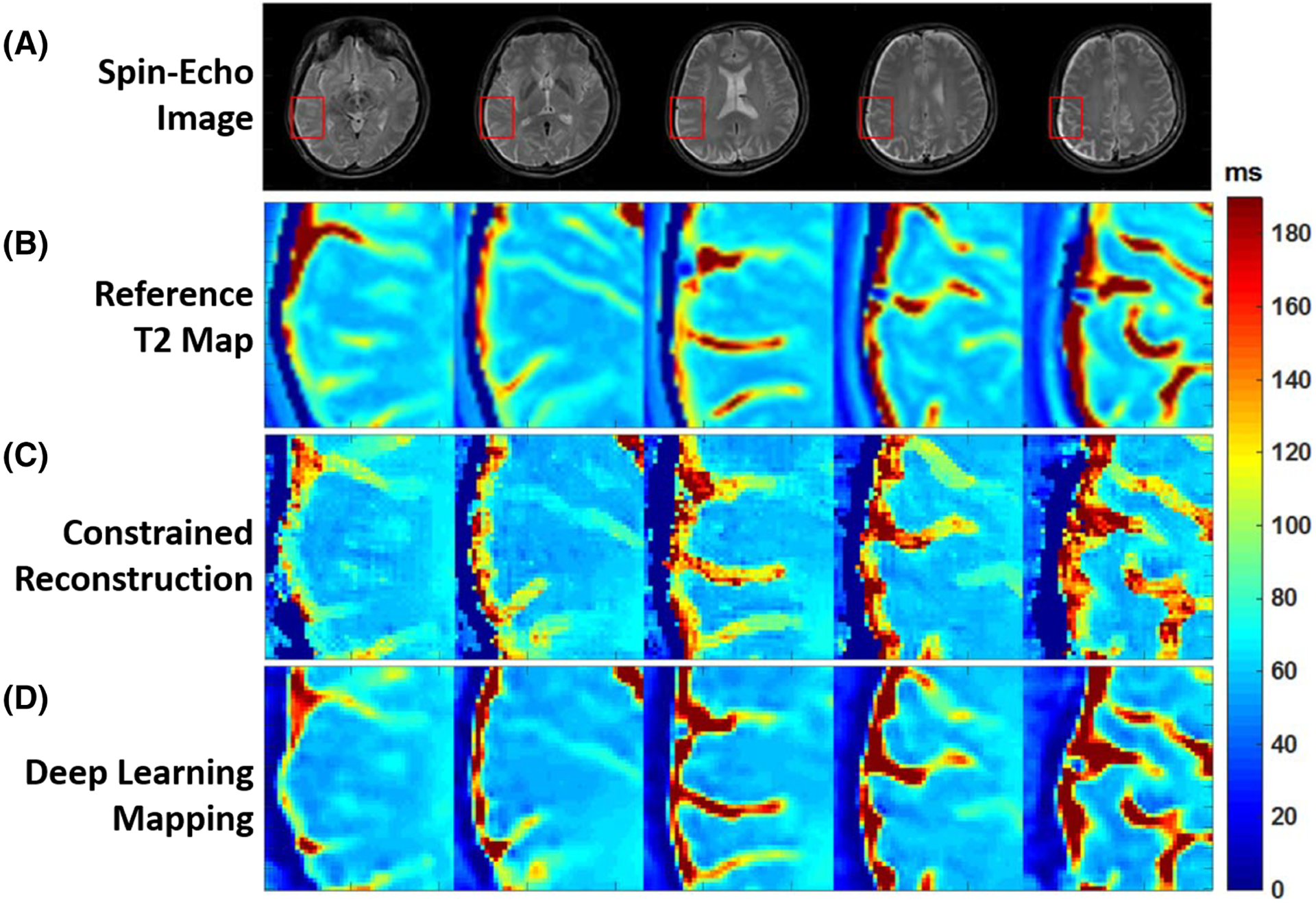
FIGURE 5.

Examples showing the comparison of T2 estimation between methods. A, Full FOV spin-echo images. B, Expanded reference T2 maps. C, Expanded T2 maps from constrained echo-detachment-based reconstruction method. D, Expanded T2 maps from ResNet. Expanded ROIs are marked by the red rectangles in A. The reconstructed T2 mappings from the echo-detachment-based method show much more noise (regular noise-like artifacts) and blurring around the texture edges. This indicates the difficulty of denoising and reducing the blurring effect at the same time for the echo-detachment-based method. However, the ResNet method simultaneously achieves both quite well, and the results show good agreement with the reference T2 mappings. (Image reproduced from Figure 6 in Cai et al. Single-shot T2 mapping using overlapping-echo detachment planar imaging and a deep convolutional neural network. Magn Reson Med. 2018. https://doi.org/10.1002/mrm.27205 with permission)
