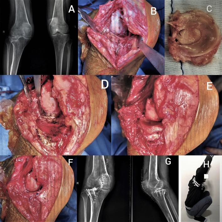
Fig. 1.
Figure showing: a Preoperative radiograph, b Large defect in lateral tibial plateau, c Lateral plateau osteo-chondro-meniscal allograft, d Tibial plateau preparation, e Fixation of allograft, f Fixation of meniscus with peripheral sutures and soft tissue closure, g Postoperative radiograph, h Postoperative clinical picture showing full range of knee motion

