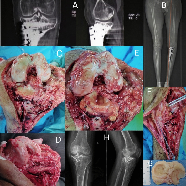
Fig. 3.
Figure showing: a Preoperative computed tomography Showing decreased joint space with implant in situ b Preoperative computed tomography scanogram showing varus deformity in knee. c Malunited medial tibial plateau without any cartilage or meniscus. d Tibial plateau preparation. e Final reconstruction with fixation of osteo-chondro- meniscal allograft. f Quadriceps V–Y plasty being performed. g Osteo-chondro- meniscal allograft. h Postoperative radiograph showing restored level of tibial plateau with optimum alignment

