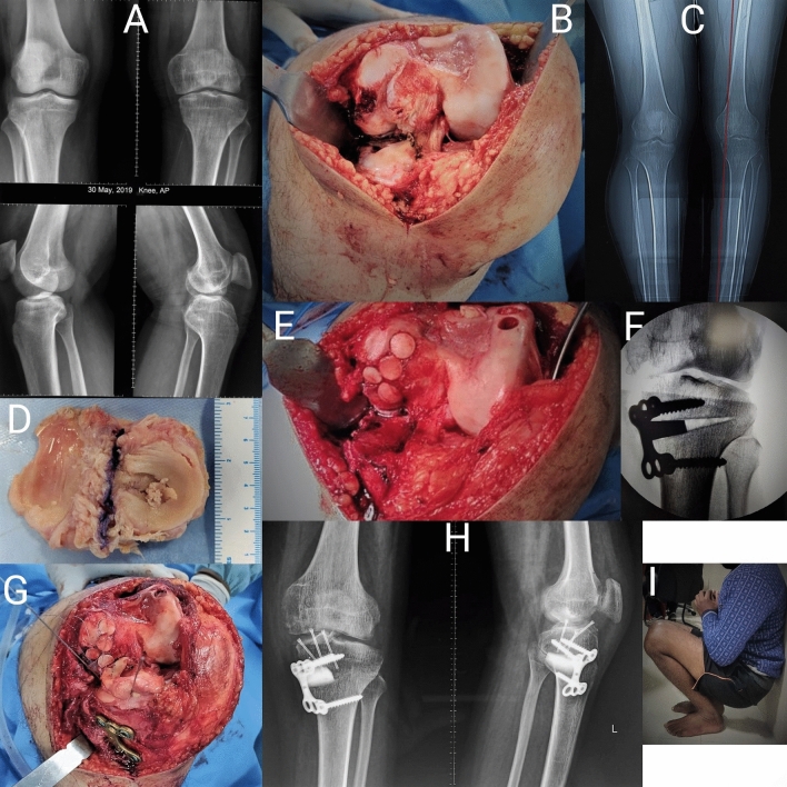
Fig. 4.
Figure showing: a Preoperative radiograph. b Kissing cartilage lesion in medial tibial plateau and femoral condyle without any meniscus. c Preoperative computed tomography scanogram showing varus deformity in the knee. d Osteo-chondro- meniscal allograft. e Tibial plateau preparation with osteochondral autologous transfer for femoral cartilage defect, f Intraoperative C-arm image of final correction after high tibial osteotomy (HTO), g Fixation of allograft with provisional K-wires after fixation of HTO with plate, (H)Postoperative clinical picture showing the full range of knee motion, i Postoperative clinical picture showing full range of knee motion

