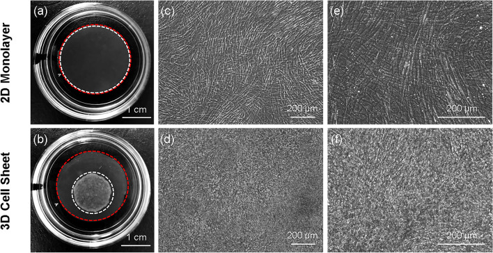
Figure 1.
Microscopic cell morphology influences macroscopic tissue structure. Macroscopic image and microscopic cell morphology of hUC-MSC 2D monolayers seeded onto an insert membrane, and contracted 3D cell sheets following temperature-detachment and placement on an insert membrane. In both groups, hUC-MSCs were seeded at 41,580 cells/cm2 initial cell densities. Macroscopic images of a (a) 2D monolayer (white dashed circle) on an insert membrane (red dashed circle, 24 mm diameter) and a (b) 3D cell sheet (white dashed circle) on an insert membrane, placed in the center of tissue-culture plastic dishes (35-mm diameter) for imaging. Morphology of hUC-MSCs in a 2D monolayer at (c) × 10 and (e) × 20 magnification, and in a 3D cell sheet at (d) × 10 and (f) × 20 magnification, observed using phase-contrast microscopy. Scale bars = 1 cm in (a) and (b). Scale bars = 200 μm in (c) through (f).

