Figure 3.
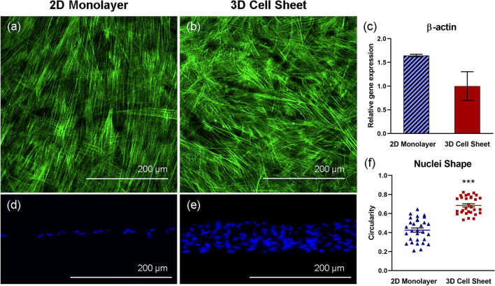
hUC-MSC actin structure changes in response to cell sheet contraction. Representative immunofluorescent images of hUC-MSC cytoskeleton (F-actin, green), imaged top-down, within (a) adherent 2D monolayers and (b) detached contracted 3D cell sheets. Cytoskeletal remodeling in response to cell sheet contraction is demonstrated by non-significant differences in (c) β-actin gene expression in hUC-MSCs in 2D monolayers and 3D cell sheets. hUC-MSC nuclei (DAPI, blue), imaged in cross-section, are elongated within the aligned cytoskeletal structure in (d) 2D monolayers and are significantly more rounded within the 3D cytoskeletal organization in (e) 3D cell sheets, evidenced by (f) nuclei circularity quantification. Scale bars = 200 μm. Values are means ± SE (n = 30: ***p < 0.001).

