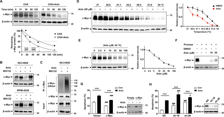
Fig. 5. Anlotinib directly targets c-Myc and promotes its ubiquitin proteasomal degradation.
A NCI-H929 cells were treated with CHX (10 μg/ml) in the presence or absence of anlotinib (5 μM) for the indicated time. The levels of c-Myc protein were evaluated and plotted in the graph. B NCI-H929 and RPMI-8226 cells were treated with either anlotinib (5 μM) and/or MG132 (5 μM) for 6 h, and then the c-Myc protein was assessed by western blotting. C NCI-H929 cells were pretreated with 5 μM MG132 for 3 h and then incubated with 5 μM anlotinib for 3 h. The cell lysates were immunoprecipitated with anti-c-Myc antibody and then probed with anti-ubiquitin antibody. D, E The binding between anlotinib and c-Myc protein was examined by the CETSA method at different temperatures or doses. The indicated proteins were evaluated by western blotting (left). CETSA curves of c-Myc were determined in the absence and presence of anlotinib. Each band intensity of c-Myc in DMSO or anlotinib group was normalized with respect to that obtained at the lowest temperature or dose (right). F Whole cell lysates of NCI-H929 cells were incubated with anlotinib followed by digestion with pronase according to the “Materials and methods” section. Then, the degree of c-Myc degradation was determined by western blot. G, H Overexpression and shRNA knockdown efficiency of c-Myc in NCI-H929 cells was measured by western blotting analysis. The effects of c-Myc knockdown or overexpression on cell apoptosis were evaluated by flow cytometry. The presented columns are given as the means ± SD. Statistically significant values compared with the DMSO group or empty vector group are depicted by * or #, respectively. ***P < 0.001, #P < 0.05, and ###P < 0.001. Con control group, Anlo anlotinib group. Data are shown as mean ± SD and representative of three independent experiments.

