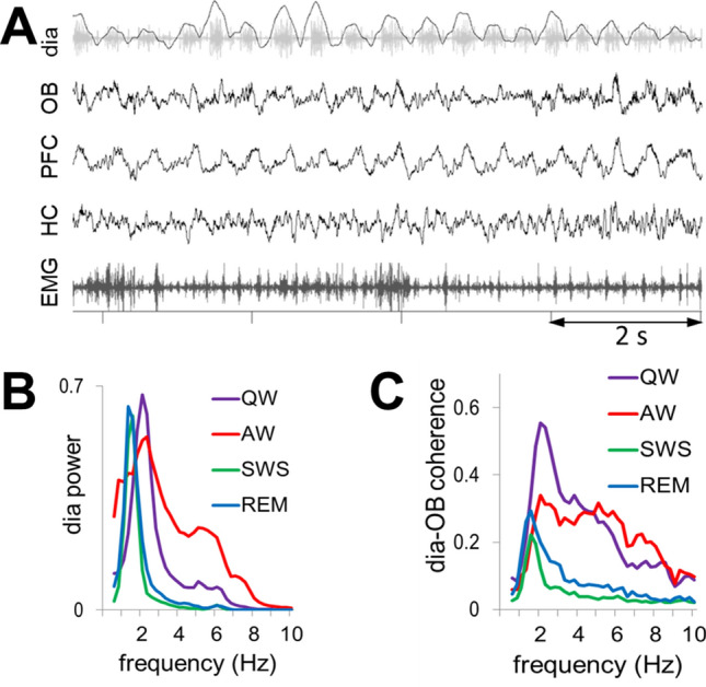
Figure 1.

(A) Sample recording of respiratory rhythm (black, inspiration up) derived from diaphragmal EMG (gray) along with LFPs in OB, PFC, and HC and neck muscle EMG in QW state. (B) Group averages of dia autospectra in different states. Note narrow RRO peaks in all recordings at ~ 2 Hz. Power is shown in arbitrary units after normalization of autospectra in individual recordings setting maxima equal to 1. (C) Group averages of dia-OB coherence spectra in different states. Note coherence peaks constrained to RRO frequencies (i.e. dia spectral peaks) in sleep and in a wider range, up to 6 Hz in wake states. In AW, dia-OB coherence does not have a clear RRO peak on the group average due to interindividual variability of the respiratory rates (see in Fig. S2).
