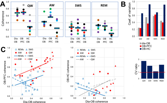
Figure 2.
Comparison of state-dependent RRO coherences in PFC and HC transferred through OB. (A) Pair-wise coherences at the respiratory frequency (RRO) between rhythmic dia activity and LFP in the OB and between OB and cortical (PFC) and hippocampal (HC) networks during different sleep–wake states. Note strong state dependence and nearly identical dia-OB and OB-PFC coherences in all recordings and considerably lower OB-HC coherence. Squares: group averages, dots: individual experiments; same colors identify individual rats across different states. (B) Variability of coherence values in individual experiments in different states. Coefficient of variation (top) and CV ratio (bottom) of coherences in the OB-HC vs. the other two signal pairs (dia-OB and OB-PFC). Note high variation of OB-HC in waking (AW and QW) and REM sleep, 2–3 times exceeding CV of the other pairs. (C) Relationship between RRO coherences connecting dia to OB C(Dia-OB) and those connecting OB to neural networks of PFC (left) and HC (right) in different states. Trend lines with nonsignificant correlations (p > 0.1) are shown in gray; solid lines show theta, and dashed lines show non-theta states. Note the significant positive correlation between dia-OB and OB-PFC but no positive correlation between dia-OB and OB-HC coherences (only a non-significant trend in waking).

