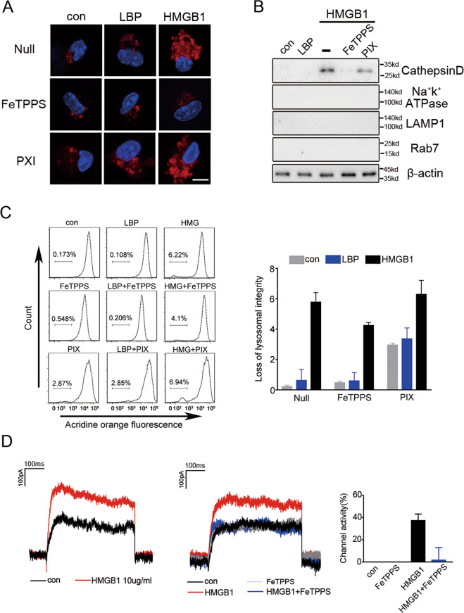
Fig. 6. FeTPPS inhibits the cytosolic delivery of LPS through preventing HMGB1-induced lysosomal rupture.
A Confocal microscopy of mouse peritoneal macrophages incubated for 4 h with DQ ovalbumin (10 μg/mL; red) alone or together with LPS-binding protein (10 μg/mL), or together with HMGB1(10 μg/mL) in the absence or presence of FeTPPS (1 μM) or PIX (1 μM) for 4 h, then stained with DAPI (blue). Scale bar: 10 μm. B Western blots for cathepsin D, Na+-K+-ATPase, Lamp1, Rab7 in the cytosolic fraction from vehicle-treated or LBP (5 μg/mL) or HMGB1 (5 μg/mL) with or without FeTPPS(1 μM) or PIX (1 μM)-treated mouse peritoneal macrophages. C Flow cytometry of mouse peritoneal macrophages stained with acridine orange and then treated for 4 h with LBP (10 μg/mL) or HMGB1 (10 μg/mL) with or without FeTPPS (1 μM) Numbers above bracketed lines indicate percent cells with loss of lysosomal staining with acridine orange (excitation, 488 nm; emission, 650–690 nm). D Whole-cell patch-clamp recording of HMGB1 in the absence or presence of FeTPPS (1 μM) induced inward current across the cytoplasmic membrane in proximity to the patch-clamp of HEK293 cells at acidic conditions (pH = 5.0). Graphs show the mean ± SD of technical replicates and are representative of at least three independent experiments.

