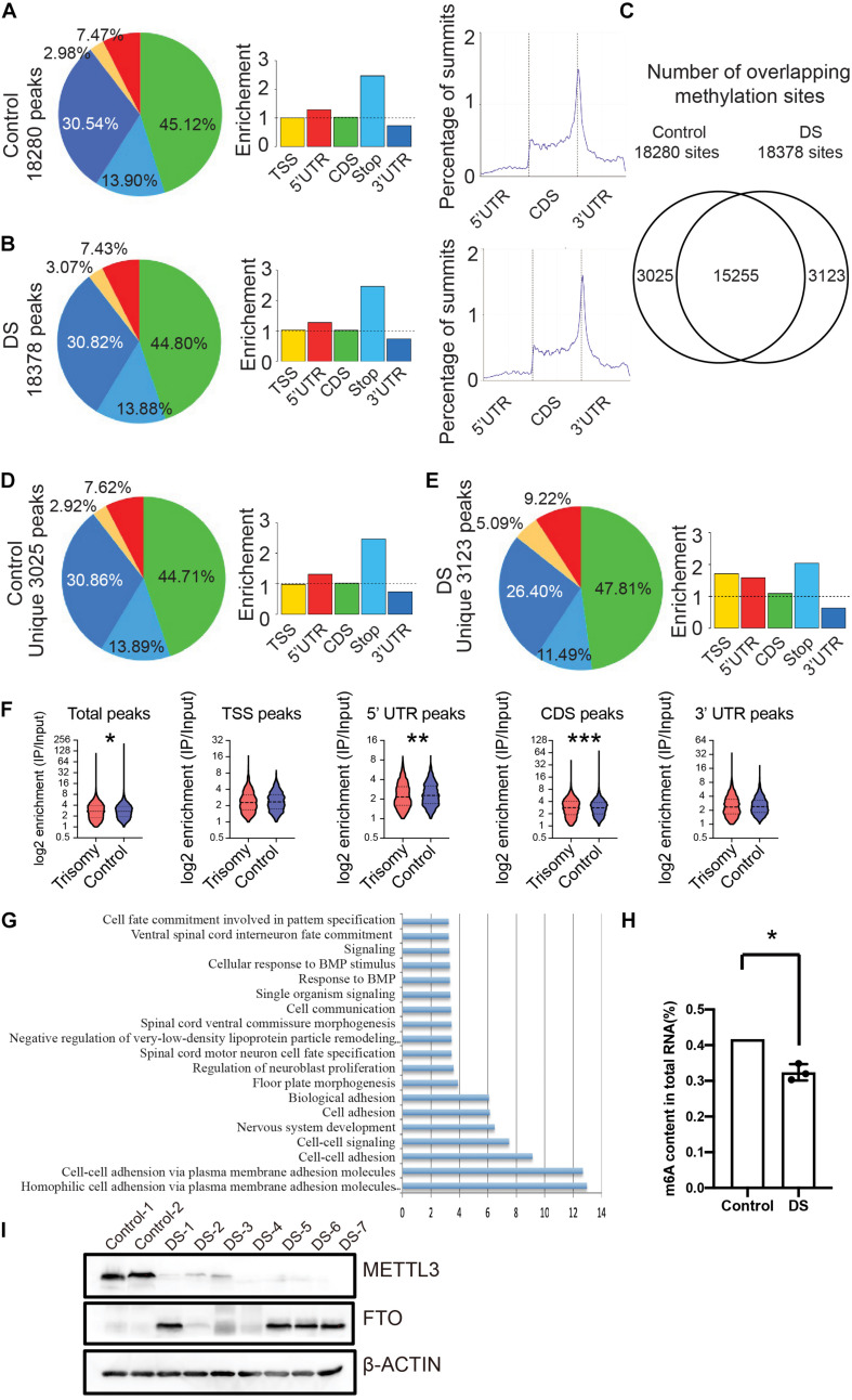
FIGURE 1.
RNA m6A modification globally decreased in fetal cerebral cortex tissues of DS. (A) Distribution of m6A peaks identified by MeRIP-seq across the length of mRNA transcripts in control cerebral cortex samples. (B) Distribution of m6A peaks across the length of mRNA transcripts in cerebral cortex of DS samples. (C) Number of overlapping m6A sites on DS and control cerebral cortex RNAs. (D) Distribution of unique m6A peaks in control samples across the length of mRNA transcripts. (E) Distribution of unique m6A peaks in DS samples across the length of mRNA transcripts. (F) Violin plot depicting the transcripts containing m6A peaks on 5′ UTR was reduced in DS compare to control subjects. (G) GO analysis of differential m6A modified genes. (H) Decrease of global m6A level in total RNA isolated from DS fetal cerebral cortex compared with control via an m6A enzyme-linked immunosorbent assay kit. (I) Western blotting analysis of METTL3 and FTO in fetal brain tissues of seven DSs and two controls. The GAPDH was used as an internal control. *p < 0.05; **p < 0.01; ***p < 0.001 compared with the control group.

