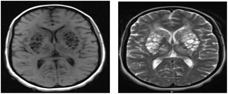
Figure 1.
a) Axial T1 weighted MRI showing bilateral multiple hyporintense cystic lesions in basal ganglia region having mass effect on adjacent ventricles by making it like a slit; b) Axial T2 weighted MRI showing bilateral multiple hyporintense cystic lesions in basal ganglia region having mass effect on adjacent ventricles making it slit

