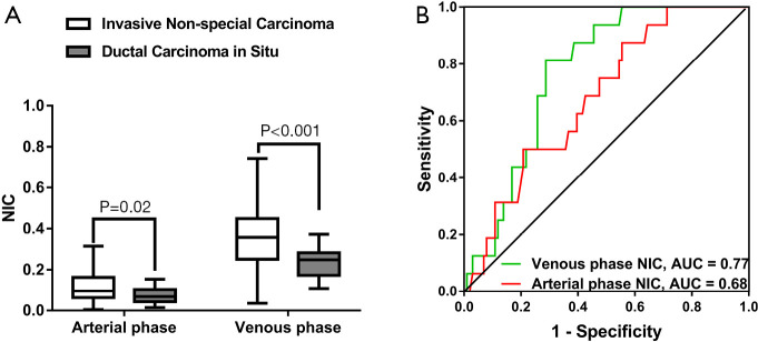
Figure 5.
Differentiation between invasive non-special breast carcinoma and ductal carcinoma in situ (DCIS). (A) Box-and-whisker plots presenting the distribution of arterial and venous phase NIC between invasive non-special carcinoma (n=101) and DCIS (n=16) of the breast. Welch’s t-test was used. (B) ROC curves of the DECT quantitative parameters, including arterial and venous phase NIC in the differentiation of invasive non-carcinoma from DCIS of the breast. NIC, normalized iodine concentration.

