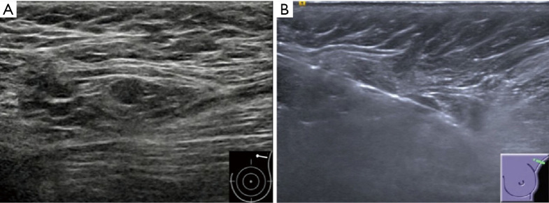
Figure 3.
Invasive ductal carcinoma in a 73-year-old woman with a negative result of axillary lymph node CNB but two lymph nodes metastases after surgery. (A) Gray-scale US image of the ipsilateral axilla shows a small (0.7×0.5 cm) suspicious lymph node with an absent fatty hilum. (B) US image obtained post-firing of CNB shows that the needle passes the lymph node precisely, but the visualization of needle may be caused by the artifact from partial volume effects. CNB, core needle biopsy; US, ultrasound.

