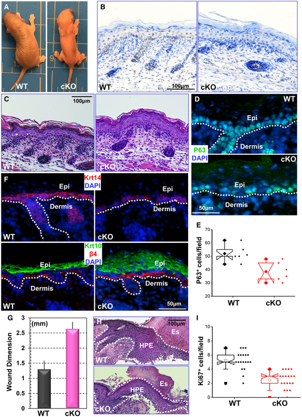
Mettl14 cKO leads to perinatal lethality. Newborn cKO pups are smaller with tight and shiny skin.
Immunohistochemistry confirms loss of Mettl14 expression in skin in cKO animals.
H/E staining of newborn skin sections from WT and Mettl14 cKO mice.
Expression of p63 in skin epidermis was examined by immunofluorescence staining in WT and cKO mice.
Number of p63‐positive cells in WT and cKO skin was quantified and shown as box and whisker plots. The plot indicates the mean (solid diamond within the box), 25th percentile (bottom line of the box), median (middle line of the box), 75th percentile (top line of the box), 5th and 95th percentile (whiskers), 1st and 99th percentile (solid triangles), and minimum and maximum measurements (solid squares). n = 6 (biological repeats), P < 0.05 (Student’s t‐test).
Skin stratification in WT and cKO skin was determined by immunofluorescence staining with different antibodies as indicated. Krt14: Keratin 14; Krt10: Keratin 10; β4: β4‐integrin; DAPI for nucleus staining. The dashed line denotes the basement membrane that separates dermis and epidermis (Epi).
Wound healing as monitored by wound size 8 days post‐injury. n = 3; P < 0.01 (Student’s t‐test). Error bar represents s.d. (standard deviation).
Histological staining of skin sections at the wound edges. Halves of wound sections are shown. Note significant reduction of HPE (hyperproliferative epidermis) in cKO skin. Es: eschar. Dotted lines denote epidermal boundaries.
Quantification of Ki67‐positive cells present in wound HPE. The plot indicates the mean (open circles within the box), 25th percentile (bottom line of the box), median (middle line of the box), 75th percentile (top line of the box), 5th and 95th percentile (whiskers), 1st and 99th percentile (solid triangles), and minimum and maximum measurements (solid squares). n = 19, P < 0.01 (Student’s t‐test).

