
Figure 1. PIF1 is important for BIR at endonuclease‐generated DSBs.
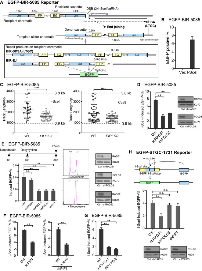
- Schematic drawing of the EGFP‐BIR‐5085 reporter and the BIR repair product by SDSA (BIR‐SDSA) or end joining (BIR‐EJ) which results in EGFP expression after splicing.
- U2OS (EGFP‐BIR‐5085) reporter cell line was infected by lentiviruses encoding endonuclease I‐SceI or empty vector, followed by puromycin selection (2 µg/ml, 2 days) and assayed for the percentage of EGFP‐positive cells by FACS analysis 4 days post‐infection.
- BIR track length was determined in the single EGFP‐positive clones derived from U2OS (EGFP‐BIR‐5085) WT and PIF1 KO reporter cell lines after I‐SceI (left) or Cas9/sgRNA (right) cleavage by sequencing the PCR products of repair junctions using genomic DNA. Group means are shown and error bars represent ± SD. Dashed lines (3.8 and 0.9 kb) indicate the upper and lower limits of track length that can be scored by this reporter.
- U2OS (EGFP‐BIR‐5085) cells expressing shRNAs for RAD51, POLD3, or shRNA vector (Ctrl) were infected by lentiviruses encoding endonuclease I‐SceI. The percentage of EGFP‐positive cells was assayed by FACS analysis 4 days later (left). Expression of RAD51 and POLD3 is shown by Western blot analysis (right).
- U2OS (EGFP‐BIR‐5085) cells carrying Tet‐On Cas9/sgRNA‐5085 (Appendix Table S1) and expressing indicated shRNA were treated by Nocodozale (0.3 μM) and 40 h later, Doxycycline (Dox, 5 μg/ml) was added. The percentage of EGFP‐positive cells was quantified by FACS analysis 48 h after induction (left). Cell cycles before and after Nocodazole treatment were analyzed by FACS following propidium iodide (PI) staining (middle). Expression of RAD51, POLD3, and RAD52 is shown by Western blot analysis (right).
- U2OS (EGFP‐BIR‐5085) cells expressing PIF1 shRNA or shRNA vector (Ctrl; left) or expressing PIF1‐WT or E307Q mutant with endogenous PIF1 depleted by shRNA (right) were infected by lentiviruses expressing I‐SceI. The percentage of EGFP‐positive cells was assayed by FACS analysis 4 days post‐infection. PIF1 expression level was determined by qPCR (Appendix Fig S3A), and the expression of PIF1‐WT or E307Q mutant is shown in Appendix Fig S3C.
- U2OS (EGFP‐BIR‐5085) cells and two PIF1 knocked‐out (KO) clones derived from the U2OS (EGFP‐BIR‐5085) cell line and generated by CRISPR KO were assayed for the percentage of EGFP‐positive cells by FACS 4 days after I‐SceI lentiviral infection.
- Schematic drawing of the EGFP‐STGC‐1731 reporter and the repair product generated by STGC is shown (top). iEGFP: internal EGFP. U2OS (EGFP‐STGC‐1731) cells expressing RAD51, POLD3, PIF1 shRNA, or shRNA vector (Ctrl) were assayed for EGFP‐positive repair events by FACS analysis 4 days post‐infection of I‐SceI lentiviruses (middle). Expression RAD51 and POLD3 is shown by Western blot analysis (bottom).
Data information: Error bars represent the standard deviation (SD) of at least three independent experiments. Significance of the differences was assayed by two‐tailed non‐paired parameters were applied in Student's t‐test. The P value is indicated as **P < 0.01, ****P < 0.0001 and n.s. (not significant) P > 0.05.
Source data are available online for this figure.