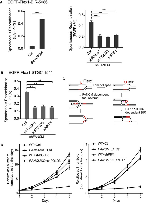
Figure 6. PIF1‐ and POLD3‐mediated BIR is important for repairing DSBs at Flex1 caused by FANCM deficiency.

- U2OS (EGFP‐Flex1‐BIR‐5086) cells were infected by lentiviruses expressing FANCM shRNA or shRNA vector (Ctrl; left) or cells from the same reporter cell line expressing shRNAs for RAD51, POLD3, and PIF1 or shRNA vector (Ctrl) were infected by FANCM shRNA lentiviral viruses (right). The percentage of EGFP‐positive cells after spontaneous recombination was quantified by FACS analysis 5 days after lentiviral infection of FANCM shRNA. Knockdown of FANCM by shRNA is shown by qPCR and Western blot in Appendix Fig S12A.
- U2OS (EGFP‐Flex1‐STGC‐1541) cells expressing shRNAs for RAD51, POLD3, and PIF1 or shRNA vector were infected by lentivirus expressing FANCM shRNA. The percentage of EGFP‐positive cells was quantified by FACS analysis 5 days after FANCM shRNA lentiviral infection. Knockdown of FANCM by shRNA is shown by qPCR and Western blot in Appendix Fig S12B.
- Proposed model for concerted roles of FANCM‐dependent fork reversal and PIF1/POLD3‐dependent BIR in protection of Flex1 stability. Pink arrow: endonuclease cleavage to remove Flex1.
- Growth curves of WT or FANCM‐KO HCT116 cells were plotted after expressing POLD3 (left) or PIF1 (right) shRNA or shRNA vector (Ctrl). Cell number was normalized to that on day one.
Data information: Error bars represent the standard deviation (SD) of at least three independent experiments. Significance of the differences was assayed by two‐tailed non‐paired parameters were applied in Student's t‐test. The P value is indicated as **P < 0.01.
