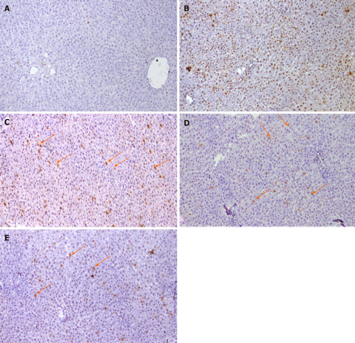
Figure 6.
Photomicrographs of liver sections Immunohistochemical stained with proliferating cell nuclear antigen. A: Negative reaction of control group; B: Diethylnitrosamine + 100 mg 2-acetylaminofluorene group showing positive stained nuclei scattered all over the field; C-E: Liver sections of rats treated with different doses of cyamidine (10, 15, 20 mg/kg) respectively, show few positive hepatocytes sporadically distributed over the field (arrow). (Magnification × 100).

