. 2021 Apr 12;54:e0850-2020. doi: 10.1590/0037-8682-0850-2020

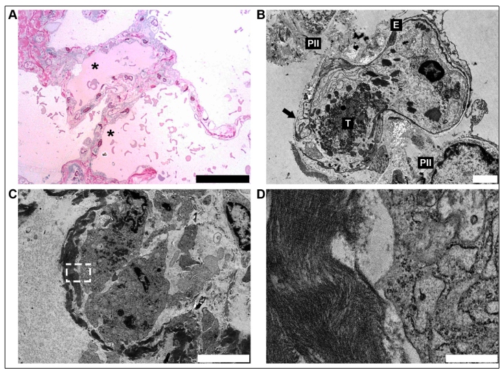
FIGURE 1: (A) Semi-thin section of the lung with an early exudative stage that was characterized principally by intra-alveolar edema (*, superior) and erythrocyte leakage (*, inferior), as well as injury to the alveolar epithelial cells (scale bar = 50 µm). (B) Electron microscopy of the pulmonary capillary showing disruption of the epithelial-endothelial barrier. Fibrin thrombi (T) inside the capillary. Type II pneumocytes (PII), endothelial cell (E) (scale bar = 2 µm). (C) Cellular debris, with nuclei in pyknosis and karyolysis of a type II pneumocyte filled by amorphous material, featuring a hyaline membrane (white, serrated edge rectangle) (scale bar = 5 µm). (D) Higher magnification of the dashed area in image C showing the fibrilar aspect of the material lining the alveoli (scale bar = 0.5 µm).

FIGURE 1: