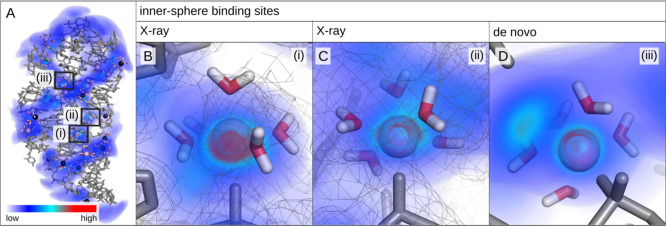
Figure 4.
Representative snapshot of the add A-riboswitch from simulations with the nanoMg parameter set and three-dimensional Mg2+ probability density. The probability density is low in the blue regions (diffusive ions) and high in the red regions (specifically bound ions). Selected inner-sphere (pink) and outer-sphere (black) Mg2+ ions are shown including the water molecules in their first hydration shell. Snapshots (i–iii) show the most probable ion-binding sites predicted from simulations with nanoMg. Snapshots (i,ii) coincide with the two inner-sphere binding sites reported in the X-ray structure.49 The experimental electron density is shown as gray mesh.

