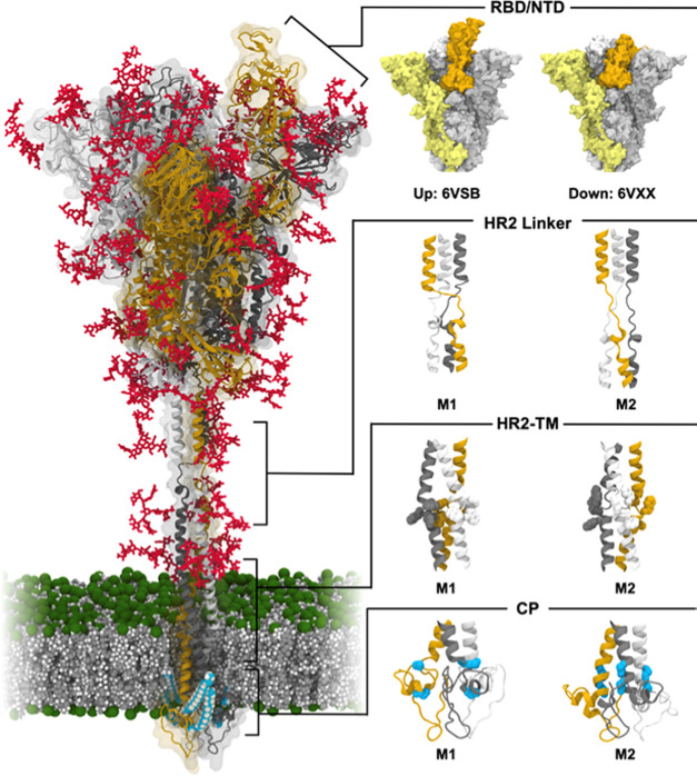
Figure 1.
Model structure of the fully glycosylated full-length SARS-CoV-2 S protein in a viral membrane. A model structure of the SARS-CoV-2 S protein is shown on the left panel. Two models for RBD/NTD, HR2 linker, HR2-TM, and CP are enlarged on the right panel. The three individual chains of the S protein are colored in yellow, gray, and white, respectively, while glycans are represented as red sticks. The palmitoylation sites of the S protein are highlighted in cyan. The phosphate, carbon, and hydrogen atoms of the viral membrane are colored in green, gray, and white, respectively. For clarity, water molecules and ions are omitted. All illustrations were created using visual molecular dynamics (VMD).30

