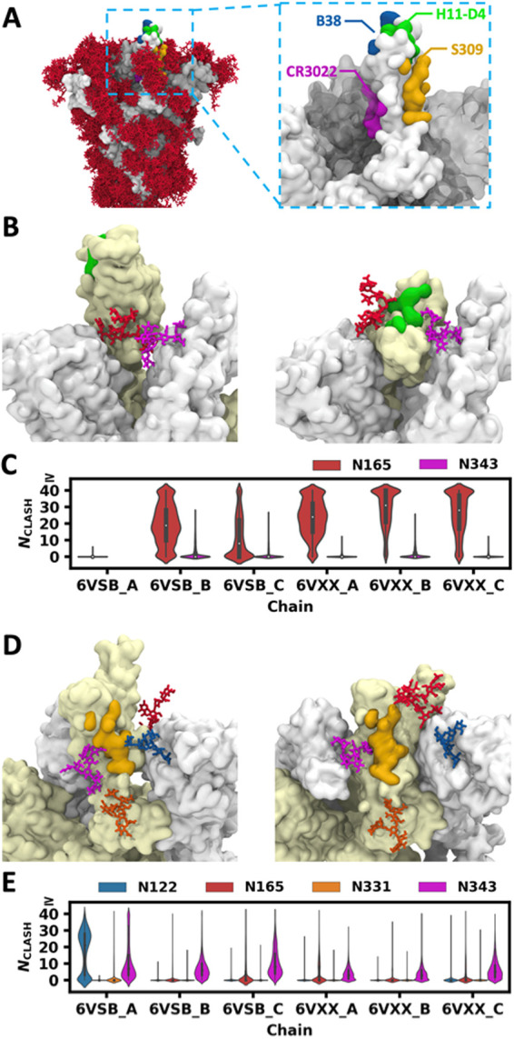
Figure 6.

Clash between glycans and superimposed antibodies. (A) Distribution of glycans when S head structures in multiple snapshots are aligned (left). Four epitopes targeted by neutralizing antibodies are shown in different colors (right). (B) H11-D4 epitope (green) in the RBD (left: open, right: closed), N165 glycan (red) on the neighboring NTD, and N343 glycan (violet) on the neighboring RBD. (C) Distributions of glycan heavy atom numbers in clash (NCLASH) with the superposed H11-D4 antibody. (D) S309 epitope (yellow) and N331 (orange) and N343 (violet) glycans on the RBD (left: open, right: closed), N122 (blue) and N165 (red) glycans on the neighboring NTD. (E) Distributions of NCLASH with the superimposed S309 antibody.
