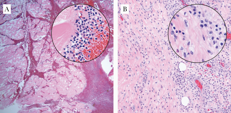
Figure 2.
Influenza A myositis and Actinomyces spp pyomyositis. (A) Photomicrograph of the lower extremity of a 35-year-old female with influenza A infection (×40, hematoxylin and eosin [H&E]; inset: ×1000, H&E). Histologically, the muscle cells are devoid of nuclei, and there is an interstitial infiltrate composed of acute and chronic inflammatory cells. (B) Photomicrograph of the rectus muscle of an 80-year-old male with an intramuscular abscess, the culture of which grew Actinomyces spp (×100, H&E; inset: ×1000, H&E). Histologically, the muscle fibers are infiltrated by acute and chronic inflammatory cells.

