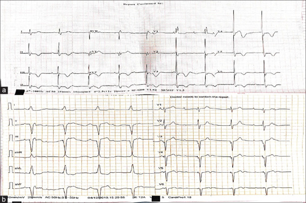
Figure 3.
(a) 12-lead electrocardiogram of the patient with a pacemaker in situ showing positive T-wave in leads I and aVL and negative T-wave in leads II, III, aVF, and V4 to V6 with precordial T-wave inversion more than T-wave inversion in inferior leads suggestive of memory T-waves. (b) 12-lead electrocardiogram of the same patient showing regular wide complex rhythm at a rate of 50 beats/min with pacer spikes suggestive of pacemaker rhythm. (Note: The lower rate of pacemaker is set at 50 beats/min to prolong the life of pacemaker)

