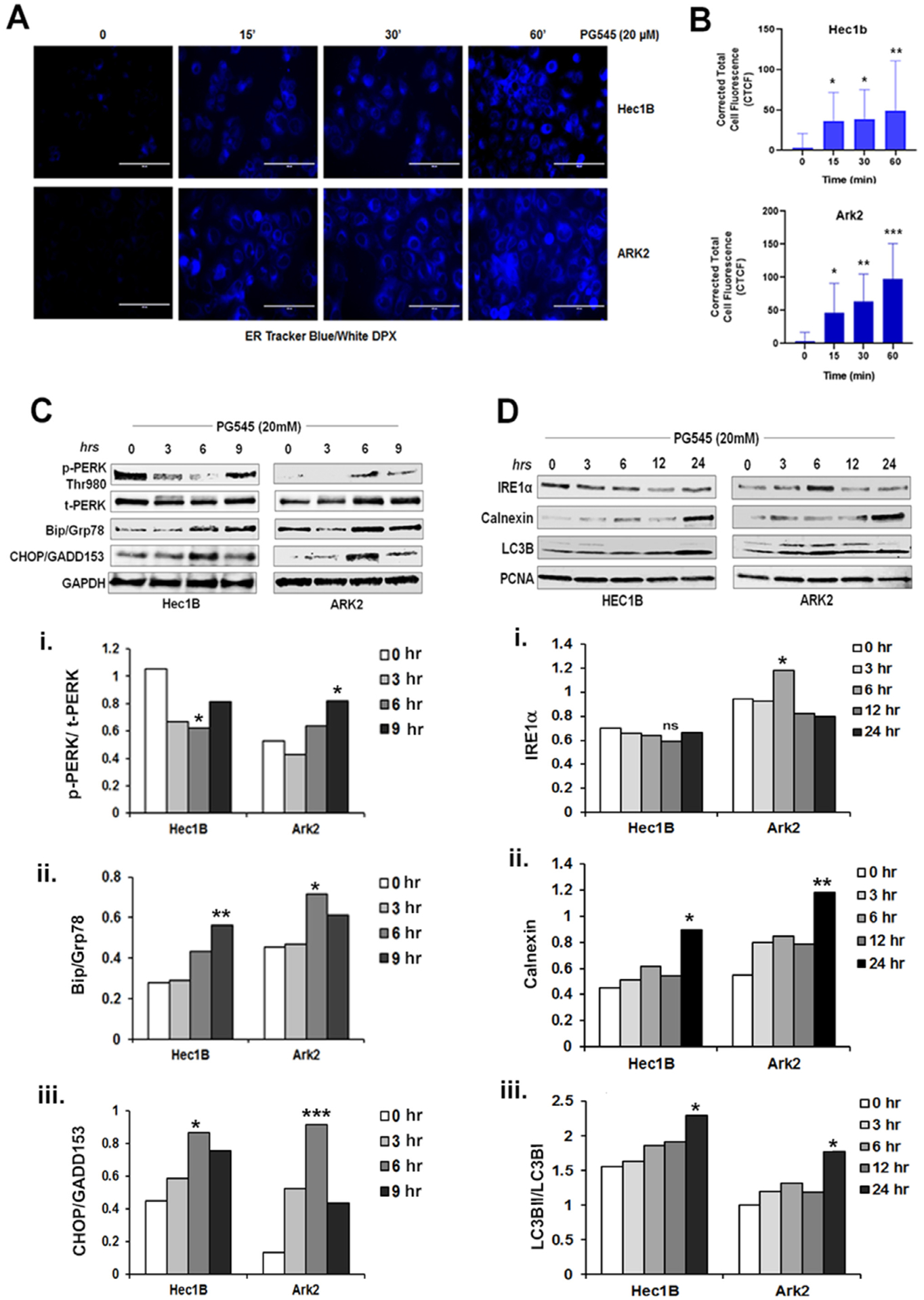
Fig. 5.

PG545 (20 μM, 0–60 mins) escalates ER activity as measured in blue-white DPX staining in Hec1B and ARK2 cells (A). Cells were incubated in Hanks balances salt solution and incubated with PG545 as mentioned in the materials and methods section. The corrected total cell fluorescence was shown in B, P values: * < 0.05, ** < 0.01. C-D. PG545 treated (20 μM) cell lysate was analyzed by Western blot using ER stress marker antibodies to p-PERK (Thr980), t-PERK, Bip/Grp78, CHOP/GADD153, GAPDH, IRE1α, Calnexin, LC3B and PCNA.
