Figure 3.
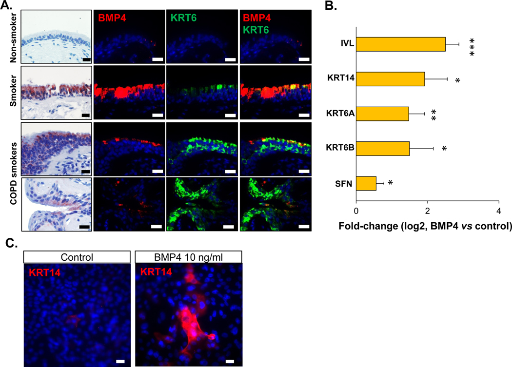
BMP4 induction of differentiation toward squamous cells. A. From left to right: 1st column - representative BMP4 immunohistochemistry staining (positive cells are shown in red), 2nd column - immunofluorescence staining of BMP4 (red), 3rd column - immunofluorescence staining of squamous cell marker keratin 6 (KRT6, green), 4th column – overlap of 2nd and 3rd column. These staining were performed on the human airway epithelium of nonsmoker with normal morphology (1st row), asymptomatic smoker (2nd row) and COPD smokers (3rd and 4th rows) with abnormal morphology. Nuclei are stained with DAPI (blue). Scale bar - 20 μm. The biopsy images with immunohistochemistry and immunofluorescence staining in each row were from the same sample. B. TaqMan assessment of fold-change (log2) in the expression of squamous cell-related genes (IVL, KRT14, KRT6A, KRT6B and SFN) from the airway epithelium after 14 days ALI culture with BMP4 (10 ng/ml) stimulation from the basolateral side vs untreated control. * p<0.05, ** p<0.01, *** p<0.001, n=3 or 4. C. Immunofluorescence top staining of the squamous cell marker KRT14 (red) on the airway epithelium after 14 days ALI culture with BMP4 (10 ng/ml) stimulation from the basolateral side (right) vs untreated control (left). Nuclei are stained with DAPI (blue). Scale bar – 20 μm.

