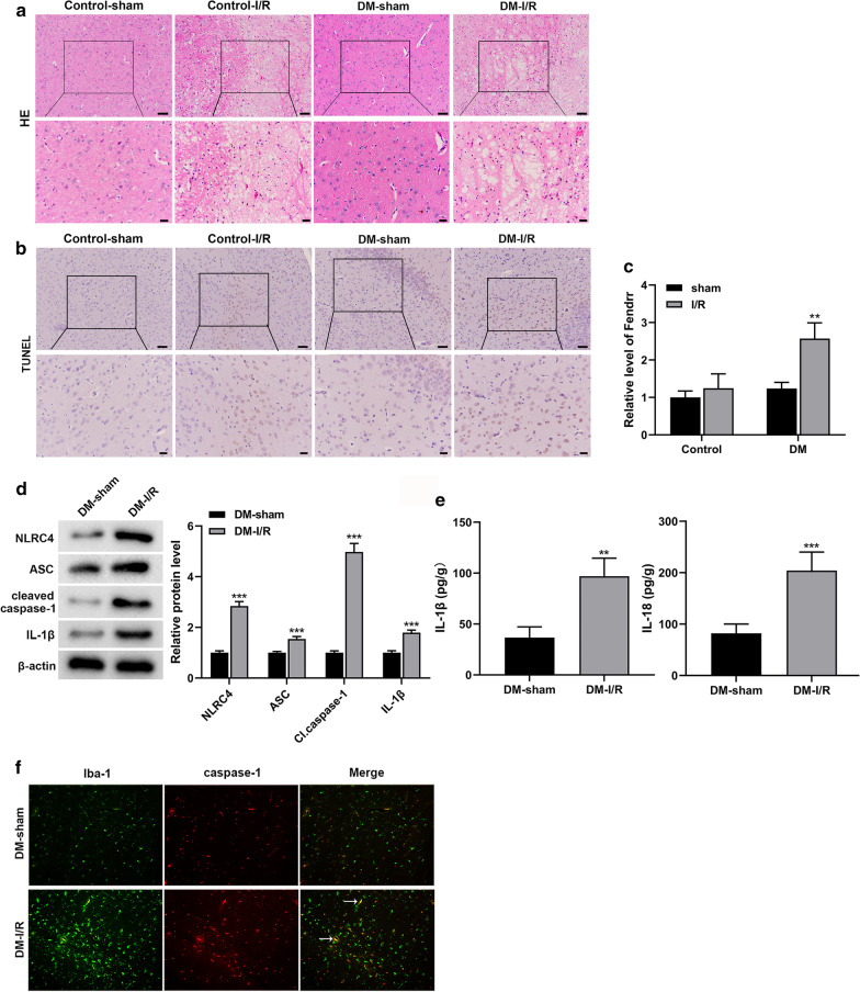
Fig. 1.
LncRNA-Fendrr and NLRC4-mediated inflammation is activated in the brain tissues of diabetic cerebral ischemia/reperfusion injury mice. The model of cerebral ischemia/reperfusion (I/R) injury in mice was established, and mice were divided into the DM-I/R group and the DM-sham group (control), n = 10/group. a HE staining was used to detect pathological changes in mouse brain tissue. Scale bar in the top line is 50 μm, and the bottom line is 20 μm. b TUNEL staining was used to detect neuronal apoptosis in mouse brain tissue. Scale bar in the top line is 50 μm, and the bottom line is 20 μm. c The expression of lncRNA-Fendrr was detected by qRT-PCR. d NLRC4, ASC, cleaved caspase-1 and IL-1β protein levels were detected by Western blotting. e The levels of IL-1β and IL-18 in brain tissue homogenate were detected by ELISA. f Detecting microglia markers Iba-1 and caspase-1 in the brain tissue of mice by Immunofluorescence (n = 3/group). Data are presented as means ± SD. P values were analyzed by Student’s t-test. **P < 0.01 and ***P < 0.001 versus the DM-sham group

