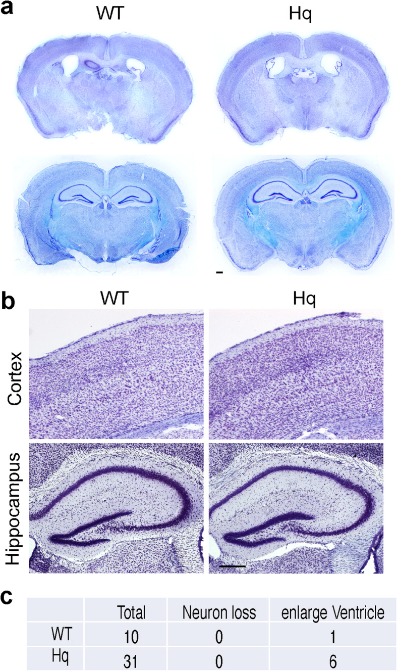
Fig. 11.

Loss of AIF in Hq mice did not obviously cause neurodegeneration in the forebrain. a Representative overview images of Hq mouse brain (P100), including somatosensory cortex, piriform cortex, thalamus and hippocampus. Nissl staining images from C57BL/6 J-Aw-J/J mice with the same genetic background were used as the control. Scale bar, 200 μm. b Nissl staining of somatosensory cortex and hippocampus from Hq and their control mice at P100. Scale bar, 200 μm. c Quantification of mouse number with either obvious neuron loss or enlarge ventricle
