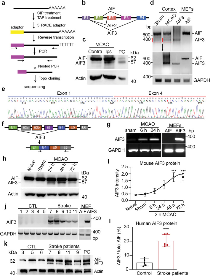
Fig. 2.
AIF3 was induced following ischemic stroke in mouse model and human patients. a The 5′-RACE scheme of AIF cDNAs from C57BL/6 mouse brain. b All three AIF isoforms identified by 5′ RACE. c Expression of AIF3 protein in C57BL/6 mouse brain 24 h after 2 h MCAO. Contra, contralateral; Ipsi, ipsilateral. PC (positive control), cerebellum lysate of AIF3 (AIFfl/Y/CamKIIα-iCre+) mice. d Expression of AIF3 mRNA in ischemic stroke mouse cortex using the common AIF/AIF3 primers (mAIF-Fw34/Fe648, Table 2). Top image is the 1st PCR with 20 cycles, middle image is the 2nd PCR using AIF3 from 1st PCR as template, and bottom image is GAPDH. e Sanger sequencing of AIF3 mRNA in ischemic stroke mouse cortex prepared from d. f Scheme for AIF3 mRNA specific primer design. g Expression of AIF3 mRNA in ischemic stroke mouse cortex using AIF3 specific primers (PCR ID: mAIF3, Table 2). MEFs were used as controls. h Expression of AIF3 protein in ischemic stroke mouse cortex at 24 h, 48 h and 72 h following 2 h MCAO determined by AIF E1 antibody. i Quantification of AIF3 protein expression in ischemic stroke mouse cortex at 6 h–72 h following 2 h MCAO. AIF3 protein levels were normalized to actin and presented as the relative intensity. Data are shown as mean ± S.E.M and analyzed for statistical significance by one-way ANOVA. n = 3. ***p < 0.001. j Expression of AIF3 mRNA in human cortex with stroke using AIF3 specific primers (PCR ID: hAIF3, Table 2). k Expression of AIF3 protein in the cortex of human stroke patients. PC, cerebellum lysate of AIF3 (AIFfl/Y/CamKIIα-iCre+) mice. l Quantification of AIF3 expression protein in human stroke patients. Data are shown as mean ± S.E.M and analyzed for statistical significance by Student t test. n = 5. ***p < 0.001

