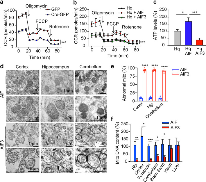
Fig. 9.
Effects of AIF and AIF3 expression on mitochondrial functions in vitro and in vivo. a OCR in AIFfl/Y/AIFfl/fl cortical neurons 3 days after GFP or Cre-GFP lentivirus infection was measured after exposure to 1 μM oligomycin, 3 μM FCCP, and 1 μM rotenone. Data are shown as mean ± S.E.M. n = 3. ***p < 0.001 by two-way ANOVA. b OCR in Hq cortical neurons 3 days after AIF or AIF3 lentiviral infection was measured by Seahorse Bioscience XF24 Extracellular Flux Analyzer after exposure to 1 μM oligomycin, 3 μM FCCP, and 1 μM rotenone. Data are shown as mean ± S.E.M. n = 3. ***p < 0.001 by two-way ANOVA. c Intracellular ATP levels were measured in Hq cortical neurons 3 days after AIF or AIF3 lentiviral infection using a luminescence ATP assay kit. Data are shown as mean ± S.E.M. n = 3. *p < 0.05, ***p < 0.001. d Mitochondrial structure of neurons in the cortex, hippocampus and cerebellum of AIF3 splicing mice (AIFfl/Y/CamKIIα-iCre+) and their littermate control AIF mice (AIFfl/Y/CamKIIα-iCre-) by electron microscopy. e Quantification of abnormal mitochondria (mito) in the cortex, hippocampus (Hip) and cerebellum of AIF3 splicing mice and their littermate control AIF mice. Data are shown as mean ± S.E.M. n = 5. ****p < 0.0001 by one-way ANOVA. f AIF3 splicing reduced mitochondrial biogenesis in brain, but not heart or liver of AIF3 splicing mice. The ratio of mitochondrial DNA relative to nuclear genomic DNA was determined by quantitative real-time PCR using primers for cytochrome b (mitochondrial) and ribosomal protein S2 (RPS2, nuclear). The data observed in AIF3 splicing mice were normalized to the littermate control AIF mice. Data are shown as mean ± S.E.M. n = 3. ***p < 0.001, **p < 0.01, *p < 0.05 by one-way ANOVA

