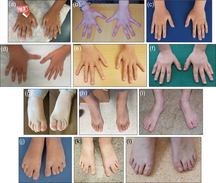
FIGURE 3.

(a) Patient 12, (b) Patient 17, (c) Patient 18, (d) Patient 19, (e) Patient 20, (f) Patient 22, (g) Patient 1, (h) Patient 12, (i) Patient 17, (j) Patient 18, (k) Patient 20, (l) Patient 22. Note the presence of fifth finger clinodactyly (d), sandal gap deformity (i, k) and cutaneous syndactyly (j, k) [Color figure can be viewed at wileyonlinelibrary.com]
