
Figure 3.
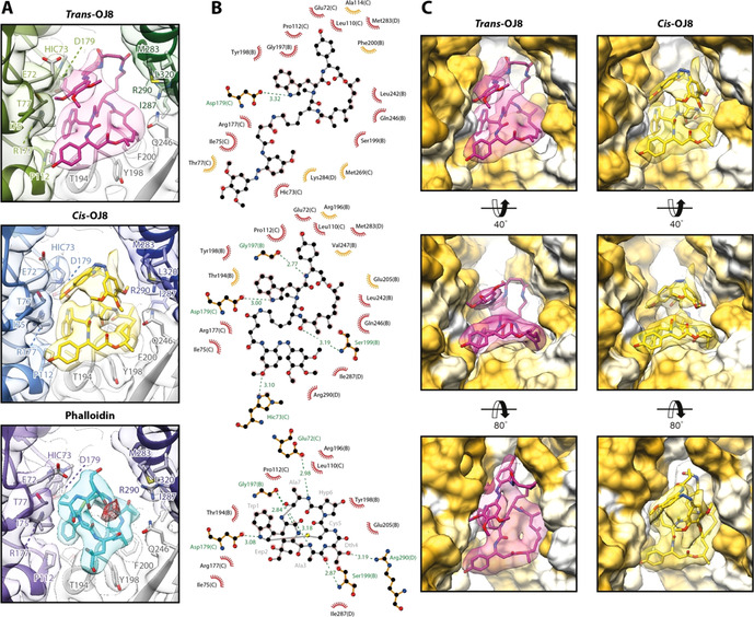
Interactions at the binding site of OJ8 in comparison to phalloidin. A) The binding site of trans‐3 (magenta) and cis‐3 (yellow) in comparison to the one of phalloidin (cyan, PDB: 6T1Y, EMDB: 10363, [9] ). Due to the scorpion tail‐like fold of the photoswitch, OJ8 resembles the three‐dimensional arrangement of phalloidin more closely than the flat conformation of JASP. The conformation of cis‐3 is remarkably similar to phalloidin (see text). B) 2D ligand‐protein interaction diagrams of the ligands shown in (A). Both hydrophobic contacts (red arcs with rays) and putative hydrogen bonds (dashed green lines) are depicted. Chain IDs are stated in brackets for protein residues. Residues that only participate in interactions with either trans‐3 (top) or cis‐3 (center) are highlighted in orange. Note that all residues involved in the binding of phalloidin (bottom), also interact with cis‐3. C) Surface representation of the OJ8 binding site from three different perspectives. While trans‐3 adopts an elongated conformation, partially filling the cavity between the actin strands (into the plane of projection), the conformation of cis‐3 is more compact, thereby increasing contacts to the subunit displayed in the upper right corner (SU D, pointed end direction), also see Movie S1.