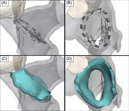
Figure 7.

Three‐dimensional rendering of the RSPV constructed using contrast enhanced CT images from a single pig. 4 placements of the circular electrode array were positioned at the ostium of the RSPV with the shape of the catheter conforming to the inner surface of the ostium (A and B). Using a conservative field threshold of 550 V/cm and uniform 3 mm tissue thickness, the field isosurfaces predict a contiguous and transmural lesion volume (green) with lesion widths ranging 3–12 mm on the outer surface of the ostium and 6.5–15 mm on the inner surface (C and D). CT, computed tomography; RSPV, right superior pulmonary vein
