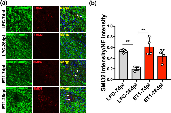
Figure 6.

Analysis of axonal dystrophy in internal capsule (IC) lesions. (a) Double immunofluorescence images of lysophosphatidylcholine (LPC)‐lesioned IC‐ and endothelin‐1 (ET1)‐lesioned IC at 7 and 28 dpl labeled with anti‐neurofilament (NF) (green) and anti‐SMI32 (red) antibodies. Nuclei were counterstained with Hoechst (blue). SMI32‐positve axons were observed in LPC lesions at 7 dpl, and ET1 lesions at 7 and 28 dpl (arrow). Scale bar, 20 µm. (b) Quantification of fluorescence intensity of SMI32 to NF (n = 4 mice/group). Mean ± SD. One‐way ANOVA, Tukey–Kramer test. **p < .01
