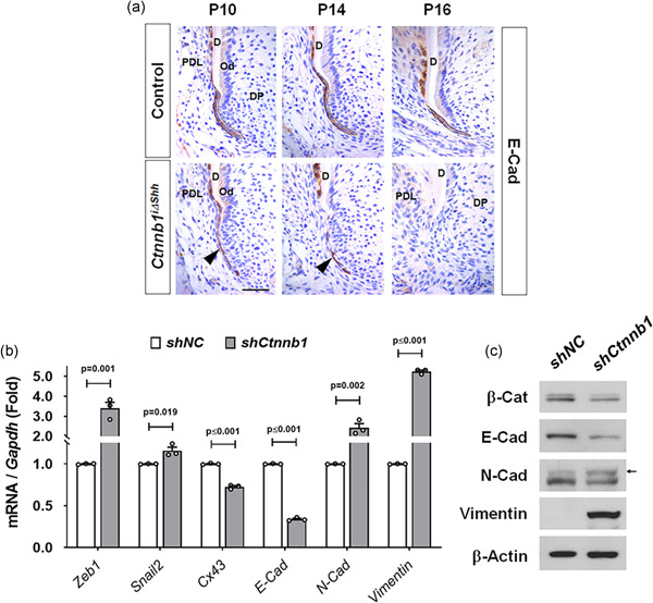
Figure 3.

Increased epithelial‐mesenchymal transition (EMT) of HERS cells by inactivation of β‐catenin. (a) Chronological changes in morphology and molecular changes in the HERS of Ctnnb1 i∆shh and control mice were compared using E‐Cad‐stained tissue sections of the mesial root of the first molar at the indicated ages after tamoxifen administration at P4. The intact HERS of the control is indicated with dotted black lines. The black arrowheads indicate the thinner and dissociated HERS of the Ctnnb1 i∆shh mice compared with those of the control. Scale bar = 20 μm. (b) mRNA transcript levels of EMT‐associated genes were analyzed by real‐time qPCR. RNA was isolated from HERS01a cells transduced with retroviruses harboring shRNA for Ctnnb1 (shCtnnb1) or the negative control (shNC). Significance was assigned for p values as indicated. (c) The protein levels were analyzed by western blot analysis. The samples were derived from the same experiment, and gels/blots were processed under the same experimental conditions. β‐Actin was used as a loading control. β‐Cat, β‐catenin. D, dentin; DP, dental papilla; HERS, Hertwig's epithelial root sheath; mRNA, messenger RNA; Od, odontoblast; PDL, periodontal ligament; qPCR, quantitative poltmerase chain reaction; shRNA, short hairpin RNA
