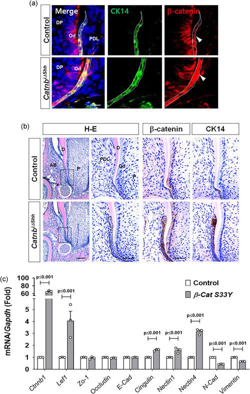
Figure 5.

Stabilization of β‐catenin in HERS results in failure of HERS dissociation. (a) Tissue sections from the mandibular first molar in the control and Catnb i∆shh mice at P12 after tamoxifen administration at P8 were stained with immunofluorescence for CK14 and β‐catenin. HERS is indicated with dotted white lines. The white arrowheads indicate that the HERS of Catnb i∆shh mice expressing CK14 shows increased expression of β‐catenin compared with the control. Scale bars = 5 μm. (b) Morphological and molecular changes in the developing tooth roots of Catnb i∆shh mice were detected with H‐E and IHC staining of the mandibular first molars at P12 after tamoxifen administration at P8. The dotted black squares in the H–E stained images indicate the magnified area of root apex, including HERS. Immunolocalization of β‐catenin and CK14 in HERS was analyzed using tissue sections from the mandibular first molars at P12. The dotted black line indicates HERS. Note the thickened and undisrupted HERS of the Catnb i∆shh mice. Scale bars = 100 μm (H–E left), 20 μm (magnified H–E and IHC). (c) mRNA transcript levels were analyzed by real‐time qPCR. RNA was isolated from HERS01a cells transfected with a plasmid driving the expression of mouse β‐catenin S33Y (β‐Cat S33Y) for the overexpression of stable β‐catenin or the control. Significance was assigned for p values as indicated. AB, alveolar bone; D, dentin; DP, dental papilla; HERS, Hertwig's epithelial root sheath; IHC, immunohistochemistry; mRNA, messenger RNA; Od, odontoblast; P, pulp; PDL, periodontal ligament; qPCR, quantitiative polymerase chain reaction
