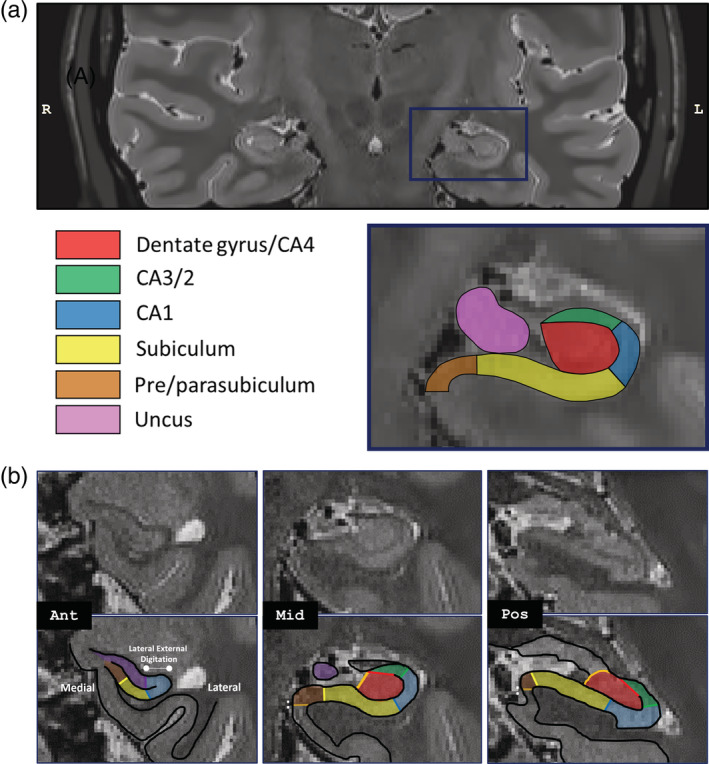
FIGURE 2.

Example hippocampal segmentation of a participant. (a) A representative high resolution (0.5 mm3) T2‐weighted coronal slice (upper panel) is displayed in native space, with a segmentation (lower panel) of the left hippocampus into its subregions, based on the protocol of Dalton et al. (2017). The displayed coronal slice is located toward the posterior end of the anterior hippocampus. Note that in native space, the right side is shown on the left. (b) The segmentation protocol is shown for the anterior, middle and posterior hippocampus without and with subregion delineations overlaid (adapted from Dalton et al., 2017). Focusing specifically on the pre/parasubiculum, this region emerges anteriorly where the lateral portion of the hippocampus bends dorsally (Ant). The lateral boundary of the pre/parasubiculum with the subiculum can be identified as a region of relatively darker gray matter on T2‐weighted images, due to dense innervations from the perforant pathway. Toward the posterior hippocampus (Mid‐Pos), there is a gradual lateral to medial shift in the location of this border. The medial border of the anterior pre/parasubiculum is located at the ventromedial edge of the hippocampus (Ant). From the appearance of the uncul sulcus onwards (Mid‐Pos), which splits the hippocampus into dorsal and ventral components, the medial border of the pre/parasubiculum occurs at the location where the medial extent of the subicular cortices turns sharply in a ventral direction (see “:”) [Color figure can be viewed at wileyonlinelibrary.com]
